38 sperm cell diagram with labels
Male Reproductive System: Labeled Diagram of Organs - Study.com The epididymis is a coiled tube present on each testicle that hosts sperm after they are produced. Sperm stored in the epididymis undergo further maturation, acquire motility, and reside there... How to draw Sperm Cell || Study of Human Spermatozoon diagram and label ... 'How to draw Sperm Cell || Study of Human Spermatozoon diagram and label the parts' is demonstrated in this video tutorial step by step.Sperm is the male rep...
Sperm Cell Labeled Diagram Stock Vector (Royalty Free) 200461103 ... Frequently used Trendsetter We're seeing significant engagement with this asset. Item ID: 200461103 Sperm Cell Labeled Diagram Formats EPS 6733 × 3563 pixels • 22.4 × 11.9 in • DPI 300 • JPG Contributor j joshya Similar images See all Assets from the same collection See all Similar video clips

Sperm cell diagram with labels
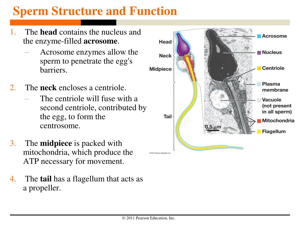
ch 8 mastering biology Flashcards | Quizlet Part A - Stages of the cell cycle Drag the labels onto the diagram to identify the stages of the cell cycle. 1. most of the cells life is spent in interphase 2. in phosphase microtubules form the mitototic spindle 3. at metaphase, the mitotic spindle is fully formed 4. in anaphase, sister chromatides separate 5. in telophase chromosomes become less condenced. Mitosis and … Draw the diagram of the human sperm and label its parts class 12 ... It is a microscopic structure, motile in nature, and fertilizes the female gamete, the egg. In mammals, the motile sperm travels with the help of a fluid known as semen. Complete step by step answer: - The human sperm can be divided into the head, the neck, the middle piece, and the tail. - The entire body is enveloped by a plasma membrane. labelled diagrams - the sperm cell labelled diagrams - the sperm cell to the right is a detailed 2D diagram of the sperm cell. there are many parts of a sperm cell. it is extremely small compared to the female egg.
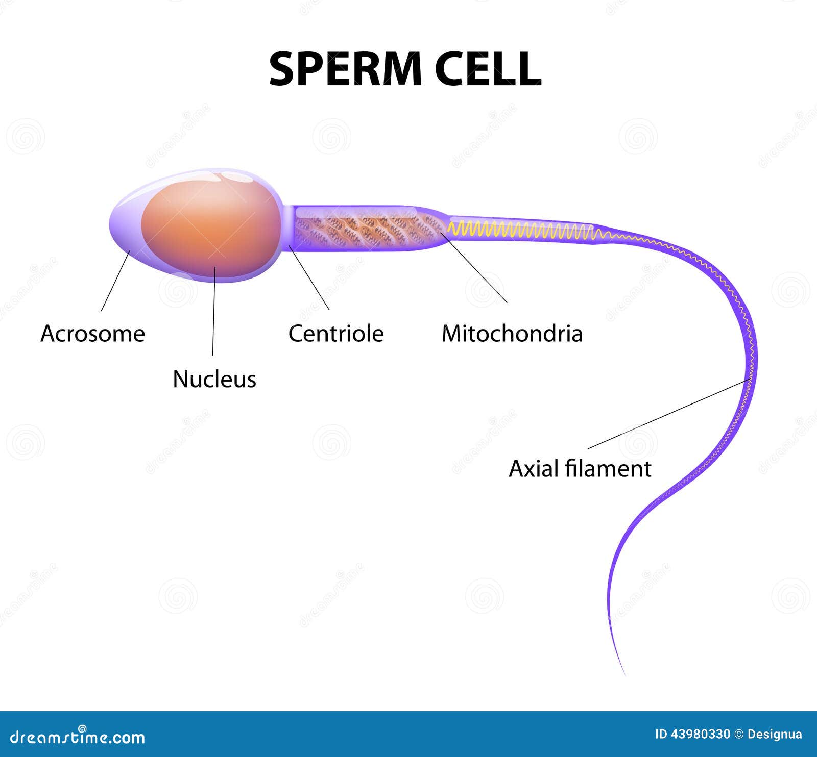
Sperm cell diagram with labels. Sperm Cells Definition, Function, Structure, Adaptations & Microscopy The head of the sperm measures 2.5 to 3.5 um in diameter and 4.0 to 5.5 um in length (um=micrometers). This results in a 1.50 to 1.70 length to width ratio They have a well-developed acrosome that covers 40 to 70 percent of the oval shaped head A slim middle section (body) that is approximately the same length as the head › cell › fulltextRecurrent inversion polymorphisms in humans associate ... - Cell May 06, 2022 · Venn diagram depicts overlap by approach for 127 tested inversions. (C–E) Evidence for single (C, 17q21) and recurrent (D, 8p23.1 [distal part chr8:8225000-8301024]; E, 11p11) loci. Left: dendrograms (centroid hierarchical clustering method) show relationships among inverted and direct-oriented haplotypes. Sperm Cells Images | Free Vectors, Stock Photos & PSD Find & Download Free Graphic Resources for Sperm Cells. 500+ Vectors, Stock Photos & PSD files. Free for commercial use High Quality Images ... Diagram showing human sex cells on white background. brgfx. 8. Like. Collect. Save. In vitro fertilization flat elements. macrovector. 22. Like. Collect. Save. In vitro fertilization concept ... Fertilization Diagram Stock Illustrations - Dreamstime Part of a flower biological diagram, vector illustration drawing with educational scheme. Labeled plant cross section with ovary, pistil, sepal and stamen. ... Sperm Cell of Human Body Anatomical Diagram. With all parts including head middle piece and tail neck mitochondrion nucleus plasma membrane for anatomy biology.
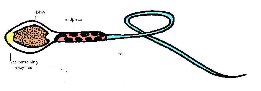
Labeled Sperm Cell Pictures, Images and Stock Photos Browse 15 labeled sperm cell stock photos and images available, or start a new search to explore more stock photos and images. Newest results Receptionist labeling sample in a laboratory Prostate labeled vector illustration. Educational male anatomy... Cell potency. From Totipotent to Pluripotent, Multipotent, and... Draw a labeled diagram of sperm. - SaralStudy Q:-With a neat diagram explain the 7-celled, 8-nucleate nature of the female gametophyte. Q:-What is oogenesis? Give a brief account of oogenesis. Q:-What is DNA fingerprinting? Mention its application. Q:-With a neat, labelled diagram, describe the parts of a typical angiosperm ovule. Q:-What is triple fusion? Where and how does it take place? An overview of sperm anatomy | Legacy Sperm is the male sex cell, also known as a gamete. Measuring approximately 0.05 millimeter (0.002 inch) long, sperm cells are made up of a few distinct parts: the tail, made up of protein fibers, which helps it "swim" toward the egg the midpiece, or body, which contains mitochondria to power the sperm's movement en.wikipedia.org › wiki › Human_penisHuman penis - Wikipedia The human penis is an external male intromittent organ that additionally serves as the urinal duct.The main parts are the root (radix); the body (corpus); and the epithelium of the penis including the shaft skin and the foreskin (prepuce) covering the glans penis.
Sperm - Wikipedia The main sperm function is to reach the ovum and fuse with it to deliver two sub-cellular structures: (i) the male pronucleus that contains the genetic material and (ii) the centrioles that are structures that help organize the microtubule cytoskeleton. Anatomy Sperm and egg fusing ( fertilisation) Cambridge Assessment International Education Cambridge … 1 The diagram shows a leaf on a plant. Sun water from the soil carbon dioxide from the air simple sugars made in the leaf Which characteristic of life is represented by this diagram? A excretion B nutrition C respiration D sensitivity 2 The diagram shows how Homo sapiens (modern people) could have evolved from earlier ancestors. Homo habilis ... Draw a labelled diagram of sperm. - Byju's Describe the various steps involved in the sexual reproduction in animals. Draw labelled diagrams to show the fertilisation of an ovum (or egg) by a sperm to form a zygote. Biology. Science for Tenth Class - Part III - Biology. Standard X. The Cell - ScienceQuiz.net The green part of the cell labelled X in the diagram is known as? a chloroplast? cytoplasm? the cell membrane? the cell nucleus ; A group of similar cells that have a shared function is known as? tissue? a system? cytoplasm? an organ; The diagram shows a group of onion cells. The parts labelled A, B and C respectively are? A = cell wall, B = cytoplasm, C = nucleus? A = cell …
What is the structure of a mature human sperm cell? - Lifeeasy Biology: Questions and Answers
Testes: Anatomy and Function, Diagram, Conditions, and Health Tips This layer is made up of Sertoli cells that aid in the production of hormones that generate sperm. Among the Sertoli cells are spermatogenic cells that divide and become spermatozoa, or sperm...

Line Diagram Of A Sperm - ClipArt Best
Recurrent inversion polymorphisms in humans associate with ... - Cell 06/05/2022 · Venn diagram depicts overlap by approach for 127 tested inversions. (C–E) Evidence for single (C, 17q21) and recurrent (D, 8p23.1 [distal part chr8:8225000-8301024]; E, 11p11) loci. Left: dendrograms (centroid hierarchical clustering method) show relationships among inverted and direct-oriented haplotypes.

Reproduction flashcards | Quizlet
Structure and parts of a sperm cell - inviTRA Structure and parts of a sperm cell 0 This labelled diagram shows the structure of a sperm cellin detail, which has the following parts: Head With its spheric shape, it consists of a large nucleus, which at the same time contains an acrosome. The nucleus contains the genetic information and 23 chromosomes.

Brenda's A & P Eportfolio: Objective 71 & 72: How sperm move and evolutionary fitness
Sperm Cell - The Definitive Guide | Biology Dictionary The sperm cell diagram below shows multiflagellate fern cells. Sperm cells from the fern plant. Most motile spermatozoa have flagella to help them swim through fluids - the seminal fluid produced by males and the mucus membranes of the female reproductive tract. Flagellum movement requires a consistent energy source.

Sperm Cell Diagram Labeled - ClipArt Best
quizlet.com › 397029425 › bisc-104-mastering-biologyBISC 104 Mastering Biology Chapter 7 Flashcards - Quizlet Discuss the following problem. Two adjacent cells in the nematode worm normally differentiate into an anchor cell (AC) and a ventral uterine precursor (VU) cell, but which of the two becomes the AC and which becomes the VU cell is completely random: the cells have an equal chance of adopting either fate, but they always adopt different fates.
PPT - Sperm Structure and Function PowerPoint Presentation, free download - ID:4130804
Sperm Diagram Stock Illustrations - 415 Sperm Diagram Stock ... Download 415 Sperm Diagram Stock Illustrations, Vectors & Clipart for FREE or amazingly low rates! ... Ocean depth zones infographic, vector illustration labeled diagram. Oceanography science educational graphic information. Depth at which sperm whales live and. ... Blue sperm cell vector illustration. 3d fertilisation isolated.

Second Questions
Spermatogenesis Diagram & Function | What is the Process of Sperm ... For example, human body cells, or somatic cells, each have 46 chromosomes. During meiosis, sperm and eggs are formed, and each of these has a haploid genome, or 23 chromosomes.

BIOLOGY CST PracticeReleased California State BiologyTest Questions. © California Department of ...
GCSE Cells | Revise Parts of Structure like Mitochondria Cell membranes allow some substances into and out of the cell whilst blocking others. It is because of this that it is said to be a semi-permeable membrane. Mitochondria are the engines of life as they provide energy from respiration. The cell wall (where present) provides support and structure for a cell and is freely permeable. All cell types ...
Structure of a sperm cell stock vector. Illustration of human - 43980330
Specialised animal cells - Cell structure - BBC Bitesize The tail enables the sperm to swim. Sperm are the smallest cells in the body and millions of them are made. Egg cell. The cytoplasm contains nutrients for the growth of the early embryo. The ...
Reproductive System Worksheet Answers - WikiEducator
Year 7 - Science Revision Guide - Chauncy School Every month an ovum (egg cell) is released from an ovary into the oviduct. This is called OVULATION. If there are sperm cells in the oviduct the ovun may join with one of them. This is called FERTILISATION. The fertilised ovum then travels down to the uterus where it grows Into a baby. The diagram below shows what happens to the ovun after it is

Sperm Cells for Artificial Reproduction and Germ Cell Transplantation - European Urology Supplements
Animal Cells: Labelled Diagram, Definitions, and Structure The endoplasmic reticulum (s) are organelles that create a network of membranes that transport substances around the cell. They have phospholipid bilayers. There are two types of ER: the rough ER, and the smooth ER. The rough endoplasmic reticulum is rough because it has ribosomes (which is explained below) attached to it.
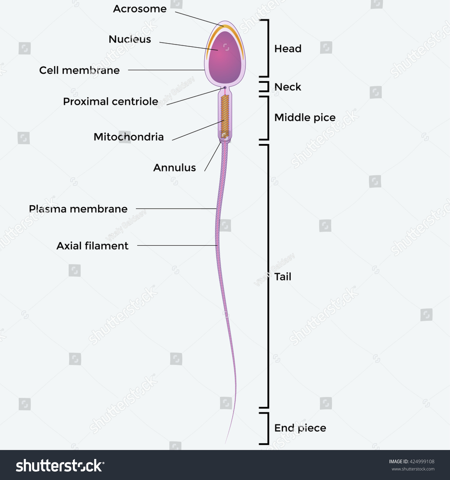
Illustartion Showing Structure Sperm Cell Stock Vector 424999108 - Shutterstock
› articles › s41586/020/2157-4Construction of a human cell landscape at single-cell level Mar 25, 2020 · a, Illustration of the experimental workflow using the microwell-seq platform.b, t-SNE analysis of 599,926 single cells from the HCL.Differentiated cell culture data and granulocyte-colony ...

Sperm Cell Diagram Labeled - ClipArt Best
Pearson Edexcel International GCSE Tuesday 8 January 2019 09/01/2019 · (ii) In the scientists’ method, sperm cells containing X chromosomes are separated from sperm cells containing Y chromosomes. This is their method. • add sperm cells to a solution of dye that stains DNA, so that the DNA reflects light • pass the sperm cells through a cell sorter • shine light on the sperm cells as they pass through the ...

Organelles of sperm
AP Biology Unit 4 Chapter 12 Flashcards | Quizlet Drag the labels onto the chromosome diagram to identify the locations of and distances between the genes. Gene m has already been placed on the linkage map. a. 12 cm b. 5 cm c. d d. p. Drag the terms to the appropriate blanks to complete the sentences below. Not all terms will be used. 1. In the P generation, a true-breeding pea plant with genotype YYRR is crossed with a true …
Post a Comment for "38 sperm cell diagram with labels"